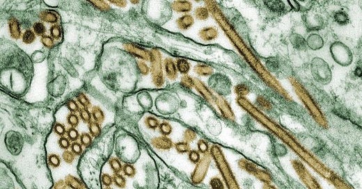
Here, photo of Avian Flu proves the only place infected with Avian flu is Wikistan. The file is from the CDC, but surprise, there is no associated paper or methods to describe the provenance of this photo and characterization of said particles. It looks so believable though.
So what is it this? It’s a big FAT lying Chick! If it’s a light microscope then a virus said to be 100nm is taking up large area of the cell, if it’s an EM photo, it means these cells are just a few 100 nm long? I got it, the cells are “micro chickens”! And, why don’t they count virus particles not antigens. Oh, maybe because there is no virus??
Of course the NIH will give the CDC-found virus a plug for the vaccine: https://www.nih.gov/news-events/nih-research-matters/progress-human-avian-flu-vaccine
The same authors from the CDC page HAVE written a related paper like this one
where they write:
FTE cells were infected apically with influenza virus at an MOI of 1, and the cells were fixed at 8 h p.i. and stained for viral antigen (HA or NP) and cell surface markers (tubulin or Jacalin). Cellular tropism for influenza virus infection was quantified by counting HA/tubulin- and NP/Jacalin-positive cells from infected cultures derived from four individual animals.
👉WHERE EXACTLY DID THEY GET THE INFLUENZA VIRUS TO INFECT THE FTE CELLS?
An HPAI H5N1 subtype virus, A/Vietnam/1204/2003 (VN/1203), was grown in the allantoic cavities of 10-day-old embryonated hen's eggs. Allantoic fluid was clarified by centrifugation....Virus titers were determined by plaque assay. The identity of virus genes was confirmed by sequence analysis to verify that no inadvertent mutations were present during the generation of virus stocks [WTF?].
👉Guys, where exactly did they get the HPAI H5N1 subtype virus?? Ahhh, this little proton is getting tired of typing. Lets see the viroLIEgy.com manual on how to find Flu A!
Summary from ViroLIEgy (redacted):
Throat washings in Tyrode’s solution were obtained on the second day of illness from a patient acutely ill with influenza
The material was centrifugalized at 2500 revolutions per minute for 30 minutes
The supernatant fiuid was then filtered through a graded collodion membrane of 500 mp average pore size
Mice were inoculated intranasally with culture fluid of the 5th transfer
...he failed with the first 3 attempts of toxic chick embryo goo injected directly into the nose of mice and progressively made it more toxic in each passage until he got the desired results
Francis Jr. concluded his own study with the two glaring admissions of being unable to identify a “virus” using tissue culture medium and egg membrane yet somehow he felt compelled to title the study: DIRECT ISOLATION OF A “VIRUS” IN TISSUE CULTURE MEDIUM AND ON EGG MEMBRANE [WTF?].
Unsatisfied with viroLIEgy who as usual couldn’t find a virus, I wrote one of the authors who replied that they, “ground up chick lung tissue obtained from Chick-Filet but apologized for leaving out the meat sauce before centrifuging lamenting that this was a crucial methodological error.”
👉 When all else fails you can leave it to Proton Magic to find the same conclusion each time: There is NO VIRUS!
Important update Aug, 2024:
Related from Ray:


This post inspired my article today in which I attempt to keep going in the same direction:
https://rayhorvaththesource.substack.com/p/so-far-so-good
This alleged EM image shows perfectly what it looks like when you cut through cylindrically shaped objects in a chaotic arrangement for EM microscopy. Some objects are cut horizontally lengthwise as horizontal cylinders, others are cut vertically in cross-section, so that longer, bisected tubes, apparently spherical objects and obliquely cut objects are also visible. The apparently spherical ones are explained as viruses with point and declare. However, it is completely impossible to cut each of these objects exactly at the equator in a cluster of spherical objects. This is only possible with cylindrical shapes that have no equator and therefore have the same diameter at every point where they are cut through.