Kary Mullis channeled-in from Valhalla🏄
Proton Magic sponsors séance, Kary rips into cell-cultures
Proton Magic calls séance for pcr inventor and Nobel Laureate Kary Mullis who “passed away” in 2019 to return and help humanity.
Séance mood music:
Kary:
Greetings Proton! Thanks for calling. I’ve been wanting to check-in from the beyond for a while now because of my disdain for what has happened to pcr and cell cultures. All these indirect and inferred platforms that are revered by the masses are still used today to peddle BS by virology.
Proton:
Wow Kary Mullis, in the flesh! I thought you were in a witness protection program? [proton trembles in fear]-you m..m…mean you are really d.d..dead? Besides being dead what irks you lately?
Kary:
The cells used in cultures are genetically disordered and usually not even from humans. More importantly they are not from the person suspected of having a virus so this set-up is not analogous to the natural state in the person who is ill.
If viruses could multiply ad nauseam in a person, they could surely be seen causing CPE (cytopathic effect or cellular death) under a light microscope. That alone should make us ditch the monkey kidney cell or HeLa cell pos, but CPE doesn’t isolate and characterize a virus particle even if there was one, it is only looking at cells which is not a method to separate nanometer size particles.
While CPE is said to be a surrogate marker for a virus, 👉you can’t have a surrogate marker for something never found to begin with💡 If it was already found, why are you trying to find it or see CPE in a system that results in CPE patient sample or not? I’d rather be at the surf than go around in circles, but it is a grave 🪦situation for persons that believe in this clownistry.
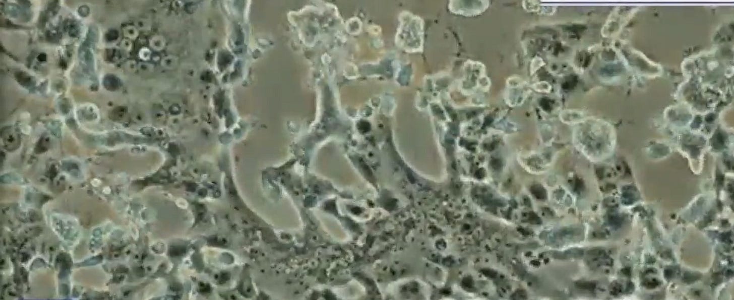
Cells dying on light microscopy. Even if they existed, virus-size particles could not be seen on light microscopy without advanced equipment (no, light microscopy cannot isolate a virus!)
Proton:
Kary first let me give an overview of the cell culture system used by virologists for the readers.
These are globs of cells that are given a sample from an ill person to supposedly see if they have a viral infection by looking for CPE. These cells are given antibiotics and antifungals ostensibly to rid the samples of bacteria or fungi that might also cause CPE, though these substances also damage cells, and would not remove other toxic material that might be in the patient sample. Not to mention the cells are starved of nutrients that also pushes CPE.
👉How can you use a test system when the system itself causes what you are looking for?
👉The so-called resistance always leaves the door open to things like, “well if there was a purified virus the test could rule it in”. That’s like looking in the mirror with your eyes that are in your head to check if you have a head. Maybe better to stick your head in a bucket of shit and wash out your brains.
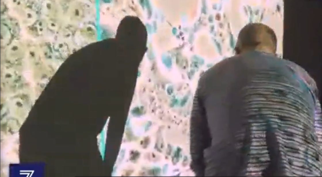
Looking at cell cultures on a big screen still doesn’t help researchers find a virus.
Kary:
I’m also dead-set against bullshit “cell culture controls”.
Proton:
We know you’re dead, but “dead-set”? Is that like the Jet-set, but you travel from grave resort to grave resort?
Kary:
Cut out the jokes, this is a grave🪦situation as I told you.
Proton:
So do cell cultures have any meaning Kary?
Kary:
No, not to rule-in or rule-out a virus.
Proton:
But what about checking cell cultures on EM for so-called “virus morphology”?
Kary:
Virologists claim then can see viruses in cell cultures on electron microscopy (EM), but EM images are just dead two-dimensional shadows on a detector plate, they are not objects that can be taken out and characterized so we don’t know what these are. I venture to say they are cell fragments and/or exosomes (vesicles from dying cells) from the CPE.
Now some have done good work to show that cell cultures with no patient sample even can find forms that look just like those that virologists claim are viruses like measles and HIV, though no virus has ever been isolated. The problem is that just showing a virus lookalike to the masses will actually make them believe there are viruses EVEN MORE SO because they will just see, “viruses”, conclude they had infected the cells in the cell culture and won’t get the logic.
Additionally, whether they are from with- or without-patient sample cell cultures, EM images are 2-dimensional dead shadows. The logic of the system itself says they can not be identified, and aside from debunking morphology as virus-finding, probably shouldn’t be done repetitively lest people claim, “see you did find viruses!”
👉How many times should we nuke a cat in a manhole to prove cats can’t withstand the blast?
Proton:
What about controls, do they fix any of the problems with cell cultures we’ve discussed?
Kary:
Wake up Proton!👉There is no such thing as a valid CPE control for cell cultures to test for CPE as due to a virus👈. For a valid control test, you would need the natural condition of THE SAME PATIENT’S sample of cells with a virus as the test sample, and for the control the SAME patient’s sample of cells without the virus in the same cell culture TAKEN AT THE SAME TIME to rule-out other possible causes of CPE in the patient that could be individual, disease, and time-dependent (like toxins, inflammation, sepsis, electrolyte disturbance for example) in order to be sure the control is exactly the same as the with-virus sample.
But, even if you could centrifuge the virus out, this makes the time stamp on the cultures change, and you have now also changed the non-virus control by shaking all the cells so that it is not a valid control anymore. Maybe the spinning would result in more or less CPE when these cells are used to test for CPE, who knows and who cares, these cells are shaken-up so they are not just the same sample sans the virus.
👉In any case you already have a virus, do you want to disprove what you have already proven to have? These two sets of conditions (same patient at the same time stamp, one with virus and one with virus removed) are clearly mutually exclusive💡
Even if we could clear all these unclearable hurdles, viruses have also been said by virologists and health authorities to be asymptomatic (Covid), or dormant (HIV). Thus negative CPE does not mean no virus was present under these narratives.
👉You can not do any rational kind of CPE study for virus finding or characterization. All you are doing is sloshing around the virus story, which is what I did in this talk too. This must stop.
Neither virologists with gotcha! smiles nor strutting an official researcher’s ID badge can find a virus. The wedding ring means gotta keep that paycheck coming.
Proton:
Ok I got it, so how can we find a virus and characterize it?
Kary:
First, be clear that “virus” only has a conceptual definition as a virus has never been found. Viruses, Bigfoot, ghosts, and many other things that cycle around conversations and texts are only constructs in the mind, there is no objective finding of these things. Maybe someday there could be but not likely, it’s more likely that persons with ulterior motives take advantage of the human nature to “believe”💡
To attempt to isolate an object that might be a virus you must first ultracentrifuge (spin) a sample taken from an ill patient looking for the 100nm size band in a gradient that separates particles based on density. The 100nm size is based on reports that claim that viruses are in that size range, but since virus particles have never actually been found there is no physical proof of a virus size. Non-virus objects that do exist like exosomes and phages are also in this size-range. Then you see if the sample is nearly purified on Electron Microscopy by confirming the objects you see from the density band are identical. Then you go back the density band you confirmed was purified in the centrifuge pellet and characterize the sample: genome, protein structure, infectivity and pathogenicity in another host, reisolate, purify, and again characterize to confirm similarity to the original. There is no cell culture involved in this procedure.
The above has never been done (probably because a particle fitting the definition of a virus has never been found), so that viruses are just guesswork and models (and sometimes fraud) based on some symptoms and lab tests that can be “positive” due to many things.
[Kary stole this blurb from here]
Proton:
Much thanks Kary, you’re not the stupid drug abusing surfer people say you are. But they really took you for a ride on pcr didn’t they?
Kary:
Proton, cool-off with all these corny Substack Posts! Get off your ass, put on some placards and go protest in front of the NIH with some crisis actors and fake news crew. Now that could get attention. So when are we going surfing? There’s some great waves here in Valhalla.
Suddenly, hissing static sound…⚡⚡⚡psss⚡…⚡...psss
Proton murmurs: Kary? Kary? Are you there? Then shouts, KARY COME BACK WE NEED YOU!!!
Proton starts shedding tears 😢
20 minutes go by, no reply. I guess the line’s dead. How sad..is Kary really dead? Proton really starts balling about Kary💧💧💧💧💧💧💧💧💧
💧💧💧💧💧💧💧💧💧💧💧💧💧
💧💧💧💧💧
Take away from today’s post:
1. Light microscopy of cell cultures that do not use cell samples from the human host being studied is not a naturalistic study and thus not applicable to the object of the study.
2. Cells used in cell cultures i.e., monkey kidney cells, have abnormal genetics which may cause them to multiply abnormally and/or die easily.
3. The procedure of cell cultures to add toxic antibiotics and starvation will itself cause CPE invalidating the procedure as study of any cause of CPE.
4. Cell cultures can not isolate nanometer-size theoretical virus particles.
5. Cell culture controls to study CPE from a theoretical virus are impossible because they would require using the same host’s cells taken at the same time and place as the test sample without a theoretical virus in it. If there was a virus, the control culture could not be a study of the same human sample sans the virus as the process of removing a virus would change the cells in the control.
6. The official narrative of dormant or asymptomatic viruses negates the ability to conclude that negative CPE means there is no virus, making the entirety of CPE study for viruses invalid.
7. The premise that CPE is a surrogate marker for a virus is untenable since no isolated virus has ever been studied to show CPE to begin with.
8. Viruses can not be isolated on electron microscopy of cell cultures as images are dead 2-dimensional shadows on a detector plate and from a mixture of multiple objects and substances of unknown content and characteristics that are impossible to study from just a 2-dimensional image.
9. Finding virus-size particles requires an ultra-centrifuge density gradient, confirmation of object morphologic purity on electron microscopy, then characterization from the centrifuge pellet. There is no cell culture involved.
For those of you who noticed, I havn’t been so nice to Kary in the past. Today though we’ve reconciled our differences:
Yours Truly,
Proton Magic & Co.
All images link to source and are for educational purposes only. The Kary Mullis conversation is fictitious., any resemblance to real persons is purely coincidental, but it could still be real if you believe it.


NOTE ON THIS POST
👉I only used Kary as a MECHANISM to make the boring topic of cell cultures readable and to get attention to the post. NOTHING about this post is about Kary or my opinion about him actually.
👉This post is also not about any other specific person, researcher, journalist, or pundit even though others have stimulated my thinking and topics inherently overlap. It is strictly on the Proton Magic take on how to conceptualize cell cultures related to virus finding and phenomena related to CPE. Study, ask questions, and search for truths without prejudice to the information provided by others.
I received a communication about genetics and exosomes.
-- On genetics, I only mentioned this, "Cells used in cell cultures i.e., monkey kidney cells, have abnormal genetics which may cause them to multiply abnormally and/or die easily."
Ref: https://pmc.ncbi.nlm.nih.gov/articles/PMC10242654/
--On exosomes I only mentioned this, "EM images are just dead two-dimensional shadows on a detector plate, they are not objects that can be taken out and characterized so we don’t know what these are. I venture to say they are cell fragments and/or exosomes (vesicles from dying cells) from the CPE."
Ref: https://pmc.ncbi.nlm.nih.gov/articles/PMC7000698/